


 21st Sep 2022
![]() Posted by Lynn Perkes on
Posted by Lynn Perkes on
Are Physical Anatomical Models Effective in Teaching Anatomy?
Teaching with Anatomical Models
The ability to see, touch, hold, and carefully examine something is an important aspect of spatial learning. The human body with its complexities and three-dimensional (3D) characteristic requires such learning. Various educational methods and learning tools are used to teach the discipline of anatomy. Learning outcomes in the study of anatomy are often related to a student’s ability to 1) demonstrate an understanding of anatomy through their proper teaching of anatomical concepts to others, 2) utilize mental 3D visualization skills to visualize structures and their relationship to other structures, 3) make and appreciate clinical correlations, and 4) demonstrate an understanding of anatomical variations and deviations. Anatomical models are excellent active learning, teaching, and demonstrating tools for those seeking to really understand human anatomy and kinesiology .
One study has shown that medical students prefer the use of cadaveric prosections paired with an active learning clinical tutorial in their study of human anatomy. ( https://pubmed.ncbi.nlm.nih.gov/34950529/)
Another study, a meta-analysis of comparative studies looking at the effectiveness of physical models in teaching anatomy, demonstrated that the use of physical models yielded significantly better results when compared to other educational methods, such as sophisticated visualization technologies, for overall knowledge outcomes, spatial knowledge acquisition, and long-retention knowledge outcome (https://pubmed.ncbi.nlm.nih.gov/26459329/). The article further stated that the physical anatomical models offer the added benefits of a low-cost alternative learning tool and the ease of use for study and accessibility .
One unique study involved veterinary students assigned to one of three teaching aid groups (anatomical physical model; textbooks; 3D computer model). The comparative efficacies of the three teaching aids were assessed through students' abilities to identify anatomical structures on MR images. Overall mean MRI assessment scores were significantly higher in students utilizing the physical model (86.39%) compared with students using textbooks (62.61%) and the 3D computer model (63.68%) (P < 0.001), with no significant difference between the textbook and 3D computer model groups (P = 0.685) (https://pubmed.ncbi.nlm.nih.gov/23349117/). Student feedback was also more positive in the physical model group compared with both the textbook and 3D computer model groups. The researchers made this concluding statement, “our results suggest that physical models may hold a significant advantage over alternative learning resources in enhancing visuospatial and 3D understanding of complex anatomical architecture, and that 3D computer models have significant limitations with regards to 3D learning.”
How to Enhance Your Learning and Teaching of Human Anatomy with Anatomical Physical Models
With the overwhelming evidence suggesting that the learning of
human anatomy is significantly enhanced by the use of physical anatomical
models,
ProHealthcareProducts.com
offers a variety of 3B Scientific anatomical models to assist with the active teaching
and learning of human anatomy. Each category of anatomical models enables
unique and hands-on demonstration of key anatomical and kinesiological
principles inherent to the musculoskeletal system. Explore below the various
anatomical model categories and learn how to use them in interactive learning
and teaching.
| Human Full Body Skeletal Models |
The adult human skeleton is comprised of 206 bones. Full body (both normal human size and mini-size) Skeletal Models provide for a good study of the skeletal system and the framework of the body in supporting and protecting organs and identifying the attachment sites for muscles that provide the levers for movement when linked to the muscular system . Full body skeletal models provide for active learning through demonstration as used in hospitals, clinics, educational settings, and laboratories. Models come in full size human adult scale or in mini versions that can be placed on table tops or handles and moved around for 3D viewing. Some models are strictly the skeletal system while others include ligaments that support the joints that they cross, display the origin and insertion sites of the muscles that produce the force for movement, allow for joint movement to demonstrate the respective movements available at the joints, and some have all of these features.
|
|

|
Human Skull Models |
The human skull is composed of two parts – the cranium and the mandible or jaw bone. These parts of the skull are made of 22 bones, 8 cranial bones and 14 facial bones, and are held together by sutures, which are synarthrodial (immoveable) joints.
- Cranial bones : frontal bone, two parietal bones, two temporal bones, occipital bone, ethmoid bone, and sphenoid bone
- Facial bones : two nasal conchae, two nasal bones, two maxilla bones, two palatine bones, two lacrimal bones, two zygomatic bones, mandible bone, vomer bone
Various skull models provide for:
- The identification of these bones as separated by sutures, or some models separate the bones by color coding the different bones
- A color scheme of muscle attachment sites with origin (red) sites and insertion (blue) sites to identify the relationship of muscles s they cross joints to produce movements of the mouth or facial expressions
- The skull sitting atop the cervical spine to show the head movements at the atlanto-occipital joint and may include the cervical spine nerves as they exit the spinal cord via the intervertebral foramen
- Facial musculature as represented by flexible and stretchable material showing the muscles of the head
| HUMAN SKULL MODES Click HERE to view all models |
|

|
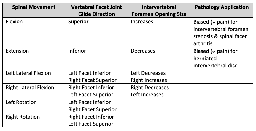
Human Spine Models |
The human spine consists of 33 individual vertebral bones that articulate with the vertebral body facets above and below. However, by the time a person becomes an adult there are only 24 separate articulating vertebrae as the sacrum and coccyx vertebrae have fused. There are 5 different spinal sections – cervical (7), thoracic (12), lumbar (5), sacrum (5), and coccyx (4).
Spine models allow for the demonstration of spinal curve
anatomy, spinal movements and the associated vertebral facet joint articulation
glide directions, and intervertebral foramen opening and closing patterns
.
This is important in the
understanding of anatomical changes related
to the intervertebral foramen opening, pressure on facet surfaces, and
compression direction on the nucleus pulposus relative to spinal curve and nerve
pressure, all of which are important considerations for understanding various
spinal pathologies and their appropriate interventions
.
| Spinal Movement | Vertebral Facet Joint Glide Direction | Intervertebral Foramen Opening Size | Pathology Application |
| Flexion | Superior | Increases |
Biased (Decreased pain) for intervertebral foramen stenosis & spinal facet arthritis |
| Extension | Inferior | Decreases |
Biased (Decreased pain) for herniated intervertebral disc |
|
Left Lateral Flexion |
Left Facet Inferior
Right Facet Superior |
Left Decreases
Right Increases |
|
|
Right Lateral Flexion |
Right Facet Inferior
Left Facet Superior |
Right Decreases
Left Increases |
|
|
Left Rotation |
Left Facet Inferior
Right Facet Superior |
|
|
|
Right Rotation |
Right Facet Inferior
Left Facet Superior |
|
|
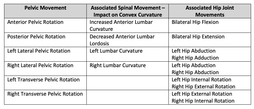
Some spine models come with an attached pelvis or pelvis and femur bone stumps. These models allow for the demonstration of pelvic movements and the associated impact (degree of convexity and concavity) to spinal curves .
| Pelvic Movement |
Associated Spinal Movement – Impact on Convex Curvature |
Associated Hip Joint Movements |
|
Anterior Pelvic Rotation |
Increased Anterior Lumbar Curvature |
Bilateral Hip Flexion |
|
Posterior Pelvic Rotation |
Decreased Anterior Lumbar Lordosis |
Bilateral Hip Extension |
|
Left Lateral Pelvic Rotation |
Left Lumbar Curvature |
Left Hip Abduction
Right Hip Adduction |
| Right Lateral Pelvic Rotation | Right Lumbar Curvature |
Left Hip Abduction
Right Hip Abduction |
| Left Transverse Pelvic Rotation |
|
Left Hip Internal Rotation
Right Hip External Rotation |
|
Right Transverse Pelvic Rotation |
|
Left Hip External Rotation
Right Hip Internal Rotation |
Spinal models present the spinal nerves as they leave the spinal cord and exit through the intervertebral foramen. Some models are specific to a respective vertebral column section for a more comprehensive study of that vertebral section’s anatomy – skeletal, neural, vascular, connective tissue . Finally, some are specific to show the pathology of a herniated or prolapsed intervertebral disc and its compression on exiting nerve roots. This aids in the teaching of dermatomes and myotomes and their used in spinal pathology diagnostics .
|
|
HUMAN SPINE MODELS Click HERE to view all models |

|
Human Joints Models |
A joint is the union or point of articulation between two or more bones. There are three major classifications of joints:
- Synarthrodial (immovable) joints such as sutures and the gomphosis of teeth
- Amphiarthrodial (slightly moveable) joints such as the symphysis pubis, intervertebral discs, costochrondral joints between the ribs and sternum, and the inferior tibiofibular union
- Diarthrodial (freely moveable) joints where most movement occurs in the skeletal system, of which there are six (6) major categories
|
Joint Classification & General Joint Name |
Planes of Motion “or”
Axes of Rotation “or” |
Anatomical Names of the Joints in the Category |
||
|
Ginglymus
(Hinge) |
One plane of motion
Uniaxial One degree of freedom |
Interphalangeal
Metacarpophalangeal of thumb Humeroulnar |
||
|
Condyloidal
(Ellipsoid) |
Two planes of motion
Biaxial Two degrees of freedom |
Metacarpophalangeal
Radiocarpal Atlantooccipital
Metatarsophalangeal
|
||
|
Trochoidal
(Pivot) |
One plane of motion
Uniaxial One degree of freedom |
Radioulnar (prox. & distal)
Atlantoaxial
Tibiofemoral (dual)
|
||
|
Enarthrodial
(Ball & Socket) |
Three planes of motion
Triaxial Three degrees of freedom |
Acetabulofemoral
Glenohumeral
Humeroradial (atypical-biaxial)
|
||
|
Sellar
(Saddle) |
Three planes of motion
Triaxial Three degrees of freedom |
Carpometacarpal of the thumb
Sternoclavicular (complex)
|
||
|
Arthrodial
(Gliding) |
Three planes of motion
Triaxial Three degrees of freedom |
|
The main diarthrodial joints of the extremities are:
- Upper Extremity : shoulder, elbow, wrist
- Lower Extremity : hip, knee, ankle
Anatomical models of individual joints allow the demonstration of osteokinematic movements, arthrokinematics motions that accompany those movements, and a demonstration of the open and close pack positions of the joints in reflecting joint congruency, ligament laxity or tautness, and the purposes for knowing this information – precautions in movements post-surgery and therapeutic intervention protocols for joint mobilizations .
| Joint (General Name, Anatomical Name, Type of Joint, Open & Close Pack Positions) | Available Osteokinematic Movements |
Accompanying “Open Chain” Arthokinematic
Motions
(Roll & Slide Directions) |
Shoulder
|
Flexion Extension Abduction Adduction Internal Rotation External Rotation Horizontal Abduction Horizontal Abduction |
Roll – anterior, Slide – posterior
Roll – posterior, Slide – anterior Roll – superior, Slide – inferior Roll – inferior, Slide – superior Roll – anterior, Slide – posterior Roll – posterior, Slide – anterior Roll – posterior, Slide – anterior Roll – anterior, Slide – posterior |
Elbow
|
Flexion Extension |
Roll – anterior, Slide – anterior
Roll – posterior, Slide – posterior |
Wrist
|
Flexion Extension Abduction Adduction |
Roll – anterior, Slide – posterior
Roll – posterior, Slide – anterior Roll – lateral, Slide – medial Roll – medial, Slide – lateral |
Hip
|
Flexion Extension Abduction Adduction Internal Rotation External Rotation |
Roll – anterior, Slide – posterior
Roll – posterior, Slide – anterior Roll – superior, Slide – inferior Roll – inferior, Slide – superior Roll – anterior, Slide – posterior Roll – posterior, Slide – anterior |
Knee
|
Flexion Extension Internal Rotation External Rotation |
Roll – posterior, Slide – posterior
Roll – anterior, Slide – anterior |
Ankle
& Foot
|
Ankle Plantarflexion Ankle Dorsiflexion Foot Inversion Foot Eversion |
Roll – posterior, Slide – anterior
Roll – anterior, Slide – posterior Slide – medial
Slide – lateral
|

| HUMAN JOINT MODELS Click HERE to view all models |
|

Related Products:
https://www.prohealthcareproducts.com/goniometers/
| Human Hand Models |
The human hand is an amazingly functional anatomical tool that is uniquely designed to help us hold or grasp objects and perform meticulous tasks. There are 27 bones in the hand (8 carpal bones, 5 metacarpals, 14 phalanges) and 27 joints (9 interphalangeal, 5 metacarpophalangeal, 5 carpometacarpal, 8 intercarpal). A hand model shows the ligamentous support to the multiple joints of the hand and fingers, and another model displays the intrinsic and extrinsic muscles of the hand . These models allow you to demonstrate and discuss some of its unique features that enhance the ability to grip and hold onto to objects . They include:
- An opposable thumb that is an enarthrodial type allowing 3 degrees of freedom or planes of motion to provide counterforce to the force of four-finger flexion
- Palmar skin that is anchored to the hand’s palmar aponeurosis via small ligamentous structures called ligamentous reticuli cutis, ensuring that held objects do not move the skin but are held firm
- The metacarpophalangeal of the thumb (only) is a ginglymus or hinge joint that only allows movement in one plane of motion ensuring a stronger opposable force in gripping
- Three natural arches; a longitudinal arch that forms the hand and fingers in flexion (gripping) and two transverse arches that allow for the 4 th and 5th fingers to “curl” around the object inferiorly
- Bifurcated
tendons of the flexor digitorum superficialis allow the tendons of the deeper
flexor digitorum profundus to continue to their distal attachment to the base
of the distal phalynx of the four fingers and serves to create a type of pulley
system configuration that enhances the gripping force of that muscle by
creating a greater mechanical advantage.
- Pes Cavus – high arch. Demonstrate the Feiss Line special test in determining a high arch and explain the potential consequences.
- Pes
Planus
– flat foot. Demonstrate the Feiss Line special test in
determining a flat foot and the potential consequences. Identify the main
ligamentous and fascia that help support the arch and explain the role of the
planta fascia in the Windlass Mechanism.
- Upper Extremity : hand, hand with radius and ulna, full arm, or full upper extremity (full arm and shoulder girdle)
- Lower Extremity : foot, foot with tibia and fibula, full leg, or full lower extremity (full leg and pelvis)
|
|
HUMAN HAND MODELS Click HERE to view all models |

Related Blog Articles:
Some Unique Anatomical Features of our Hand that Enhance our Ability to Grip and Hold Objects
Using a Handgrip Dynamometer to Teach Kinesiological Principles
Related Products:
https://www.prohealthcareproducts.com/handgrip-dynamometers/
https://www.prohealthcareproducts.com/baseline-mec...
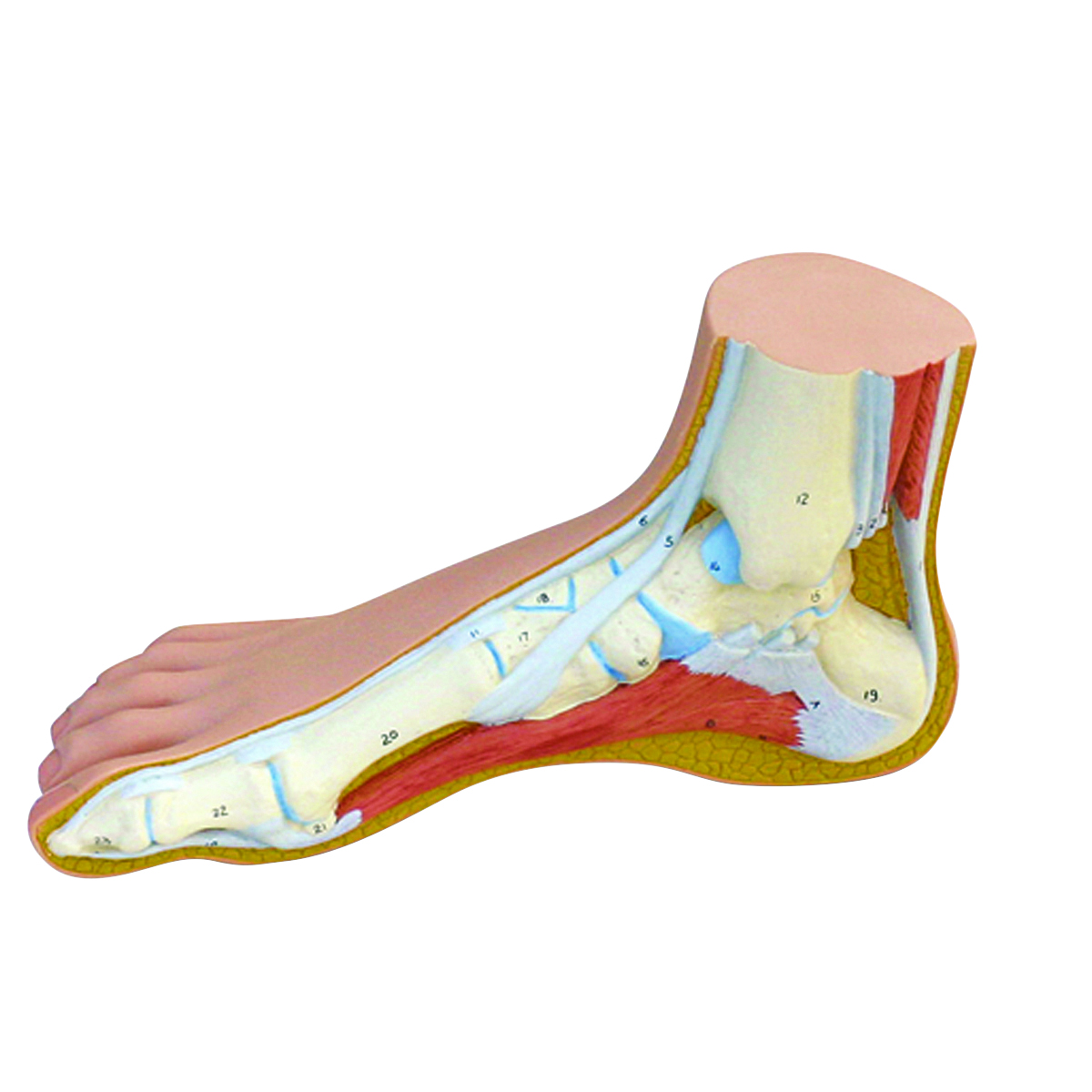
| HumanFoot Models |
The human foot has 26 bones (6 tarsal bones, 5 metatarsals, 15 phalanges) and 33 joints (9 interphalangeal, 5 metatarsophalangeal, 5 tarsometatarsal, 14 intertarsal and intermetatarsal). One foot model shows the ligamentous support to the multiple joints of the foot, toes and its arches (medial and lateral longitudinal arches and the transverse arch), and another model displays the intrinsic and extrinsic muscles of the foot and ankle allowing you to demonstrate their role in arch support, and ankle, foot, and toe movements. Other unique models are designed to demonstrate the normal and pathological arch deformities:
|
HUMAN FOOT MODELS Click HERE to view all models |
|
|
Human Leg Models |
The human leg model allows for a comprehensive review of the superficial and deep muscles of the hip and leg down to the foot as the superficial muscles can be removed to show the deeper muscles providing for a good orientation of the lower extremity musculoskeletal system . Another model specifically shows a cross-sectional view of the hip joint along with the normal anatomy showing the articulating surfaces of the hip joint for a good discussion of hip congruency (as compared to shoulder joint congruency) and the anatomical changes to the bone under conditions of osteoporosis - including common femoral fracture points due to the osteoporosis – and also osteoarthritis.
|
|
HUMAN LEG MODELS Click HERE to view all models |

|
Human Arm Models |
The human arm model allows for a comprehensive review of the superficial and deep muscles of the shoulder and arm down to the hand as the superficial muscles can be removed to show the deeper muscles providing for a good orientation of the upper extremity musculoskeletal system . The nerves and vessels of the arm are also presented . The unique design of the human arm with an enarthrodial shoulder joint proceeded by a moveable shoulder girdle supported by functional stabilizing muscles and followed by a strong hinge elbow joint, pivot forearm, and condyloidal forearm joints, and terminating in the intricacies of the hand provides a highly functional tool that can place the fingers in so many different places and work with delicate precision.
| HUMAN ARM MODELS Click HERE to view all models |
|

|
Human Pelvis Models |
The anatomical male and female pelvis models allow for a competitive analysis of the bony topography differences between the two and the purposes for which they are different. Different pelvis models enable a review of the pelvic floor musculature, the different male and female pelvic and reproductive organs, the ligamentous support, vascular, neural, and intestinal system .
|
|
HUMAN PELVIS MODELS Click HERE to view all models |

|
Human Bone Models |
The human bone models provide the skeletal components of select body parts and are made from natural anatomical casts.
| HUMAN BONE MODELS Click HERE to view all models |
|

|
Human Muscle Models |
Life size torso models and mini size full body models with removeable superficial and deep musculature allow for a full body spatial orientation of the superficial muscles of the body, as well as the inner organs behind the removeable abdominal and chest walls . A 10,000 times magnified human muscle fiber replica with neuromuscular endplate allows for a unique 3D view and analysis of a single muscle fiber cell.
|
|
HUMAN MUSCLE MODELS Click HERE to view all models |

Related Products: https://www.prohealthcareproducts.com/muscle-strength-testing-devices/
|
Human Brain Models |
Cast from a real human brain and showing the more wrinkly cortex, the neuro-anatomical brain models provide a color coordinated and removeable part configuration so that they can be disassembled to identify the major external and inner parts of the right and left parts of the human brain. The human brain is remarkable in its continuous and complex activity, capacity for storage, and individual structure based on experiences.
| HUMAN BRAIN MODELS Click HERE to view all models |
|

|
Human Eye Models |
The human eye is a very complex organ capable of distinguishing millions of different colors and shapes and its design includes over 200 unique characteristics, as compared to your fingerprint that has only about 40. It is estimated that up to 80% of learning comes through the eyes
Enlarged eye model dissects into both halves of the sclera with cornea and eye muscle attachments, both halves of the choroid with iris and retina, eye lens and vitreous humor . Another model shows the optic nerve in its natural position in the bony orbit of the eye. Helps the learner to understand how the eye constantly adjusts the amount of light that is being brought in, how it focuses on near and far objects, and produces continuous images that are instantly and rapidly transmitted to the brain for interpretation.
|
|
HUMAN EYE MODELS Click HERE to view all models |

| Ear Models |
The human ear, like many organs, is always working – constantly hearing sounds, helping with balance, self-cleaning, contain the 6 smallest bones in the human body, and are capable of detecting sound waves as low as 20Hz, and as high as 20,000 Hz. Enlarged ear models showing the outer, middle, and inner ear with their component parts and removeable bone sections enable a good demonstration of the travel of sound waves, the involvement of the eardrum, hammer, anvil, and stirrup along with the labyrinth, cochlea and auditory/balance nerve for a detailed study of the anatomy of the human ear .
| HUMAN EAR MODELS Click HERE to view all models |
|

|
Human Heart Models |
In simple terms the human heart is a pump, pumping deoxygenated blood to the lungs and oxygenated blood to organs and tissues of the body. Some interesting facts about the human heart are:
With each beat, a normal healthy heart pumps 60 to 90 milliliters (2 to 3 ounces) of blood.
Your heart pumps 1.32 to 1.58 gallons (5 to 6 liters) of blood per every minute at rest (based on a resting heart rate of 70 beats/minute).
Your heart pumps about 79 to 95 gallons of blood every hour.
Every day your heart beats about 100,800 times pumping about 1, 896 to 2,280 gallons of blood every day.
During the average lifetime, a heart pumps 55,000,000 to 66,000,000 gallons of blood.
The average adult heart weighs about 11 ounces and is the size of a person’s fist.
The average weight of an infant’s heart at birth is 20 grams or 0.7 ounces.
The coronary arteries of the adult heart are about 1/8-inch (4 mm) diameter.
Anatomical heart models with their removeable parts provide for a systematic tracing of the blood flow from its entrance to the heart via the vena cava to its exit out the aorta – being moved from chamber to chamber through open valves that separate them, and to and from the lungs before reentry to the heart carrying the newly oxygenated body in preparation for final ejection from the left ventricle to the arterial system.
|
|
HUMAN HEART MODELS Click HERE to view all models |

|
Larynx Models |
The human larynx, also called the voice box, is a hollow tubular type structure attached to the superior portion of the trachea (windpipe) that provides protection for the lower airways, aids in respiration, and plays an important role in phonation, or the production of utterance of speech sounds .
| HUMAN LARYNX MODELS Click HERE to view all models |
|

| Who is 3B Scientific and What is 3B Smart Anatomy |
Founded in 1948, 3B Scientific has grown to be one of the world’s leading manufacturers of anatomical models, medical and science equipment and education products and solutions – anatomy, skill training, healthcare and patient education. Known for their highest quality, most durable, and true-to-life product replication and simulation products for teaching and learning. 3B Scientific has as its mission to advance medical and healthcare delivery through the quality, breadth, and global reach of relevant educational and simulation products.
3B Scientific has joined forces with 3D4Medical to create an innovative and extensive virtual course library of 3B Scientific Smart Anatomy courses. By combining the advantages of highest quality hands-on 3-dimentional physical anatomical models with courses from the award-winning anatomy app Complete Anatomy, 3B Scientific customers have electronic (smart phone, tablet, or computer) free access to 11 courses with 23 lectures, 39 quizzes, and 117 different views of interactive virtual models that can be accessed anywhere for an enhanced learning experience. With this exclusive offering, customers have immediate access to 3B Smart Anatomy courses in the award-winning Complete Anatomy app and receive a free warranty extension from 3 to 5 years with every anatomy model by 3B Scientific.
|
All Human Anatomy Models |
Related Resources:
Musculoskeletal Disorders Statistics
https://www.prohealthcareproducts.com/musculoskeletal-disorders-statistics/
Arthritis Statistics